
The role of 3D cell cultures in understanding mitosis and tissue architecture
DOI:
https://doi.org/10.18054/pb.v126i1-2.32856Abstract
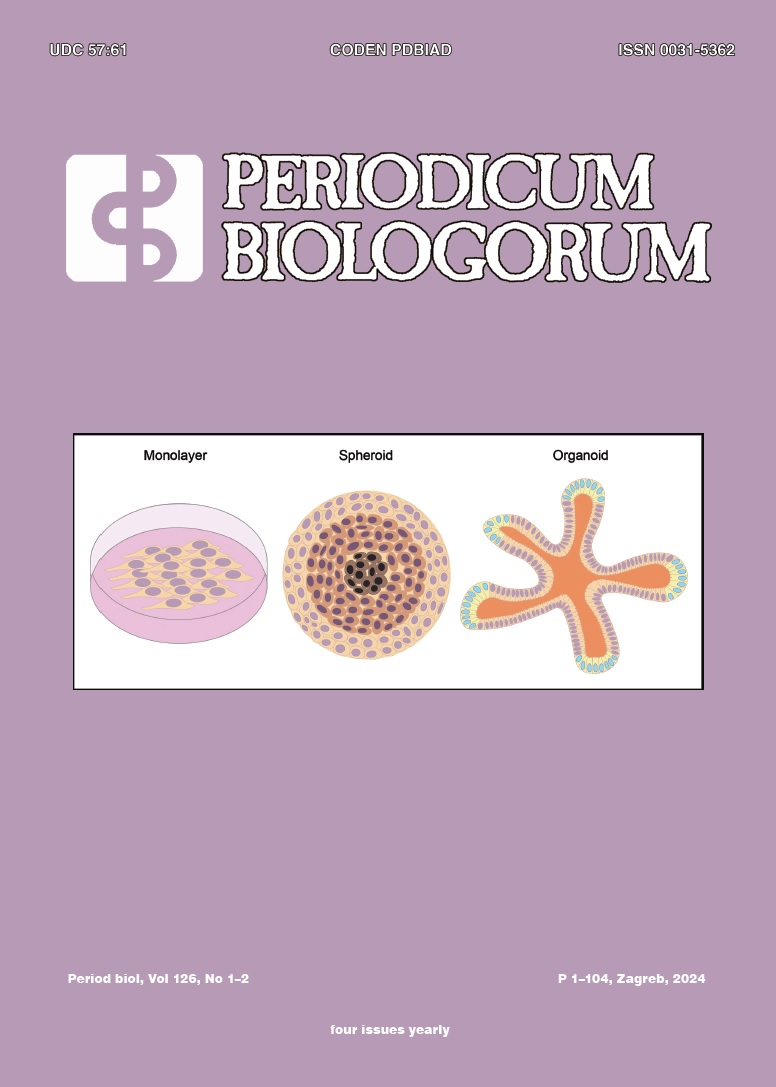
Traditional cell biology research has long relied on two-dimensional (2D) cell cultures, where cells are cultivated on flat, rigid surfaces. Although these 2D systems have contributed to significant discoveries, they often fall short in replicating the complex three-dimensional (3D) environments found in living organisms. The development of 3D cell cultures, such as spheroids and organoids, provides a more physiologically relevant model by better simulating natural tissue architecture, spatial orientation, and cellular interactions. This advancement addresses limitations of 2D cultures, including discrepancies between preclinical drug efficacy and clinical outcomes. 3D cultures exhibit greater cellular heterogeneity and altered proliferation rates, which also affects drug sensitivity and gene expression profiles. One key area where 3D cultures have shown considerable impact is in the study of mitosis—a vital cellular process for growth, development, and tissue repair—often inadequately captured by 2D models. By maintaining natural cell-environment interactions, 3D cultures facilitate a more profound understanding of mitosis and its regulation, thereby enhancing our comprehension of human biology and related diseases, including cancer.
Downloads
Published
Issue
Section
License
The contents of PERIODICUM BIOLOGORUM may be reproduced without permission provided that credit is given to the journal. It is the author’s responsibility to obtain permission to reproduce illustrations, tables, etc. from other publications.
